
Aero-TiO2 three-dimensional nanoarchitecture for photocatalytic degradation of tetracycline

The morphology of the hollow TiO2 microtetrapods is illustrated in Fig. 1. The dimensions of the arms of aero-TiO2 microtetrapods vary in the range from 20 to 40 µm in length and 1 to 3 µm in diameter. The wall thickness of the TiO2 microtubes is around 50 nm.

(a, b) SEM images of the aero-TiO2 material consisting of hollow microtetrapods, and (c) the schematic route of the aeromaterial preparation, starting with initial ZnO microtetrapods, atomic layer deposition of TiO2 on their surface (also illustrated in cross section) and, finally, removal of sacrificial ZnO substrate in the presence of HCl and H2 gases at 800 °C.
The XRD pattern shown in Fig. 2a demonstrates the presence of the rutile phase TiO2 (JCPDS 00–021-1276) and the ternary compound Zn2Ti3O8 (JCPDS 01–073-0579). All diffraction lines of the TiO2 and Zn2Ti3O8 were indexed by tetragonal TiO2 with the space group P42/mnm(136) and cubic Zn2Ti3O8 with the space group Fd-3 m(227).

XRD pattern (a) and Raman spectrum (b) of the fabricated aero-TiO2 material.
The sizes of crystallites were determined considering the main peaks of the compounds and were found to be 50 nm for rutile TiO2 and 36 nm for Zn2Ti3O8, which were determined by the Scherrer equation (Eq. 1):
$$D=0.89\lambda /\beta \text cos\theta$$
(1)
where, λ is the wavelength (Co Kα, λ = 1.7903 Å), β is the full width at the half-maximum (FWHM) and θ is the diffraction angle.
It was found previously that aero-TiO2 can be obtained in a mixture of anatase–rutile compound with Zn2TiO4 inclusions by using the same ALD process with subsequent selective wet chemical etching of ZnO sacrificial template13. Higher annealing temperature and the different approach we used for the ZnO removal allowed one to obtain a composite consisting of rutile phase TiO2 and cubic Zn2Ti3O8.
The XRD data are corroborated by the Raman scattering analysis (Fig. 2b). The rutile structure of TiO2 belongs to the space group \(D\genfrac0pt144h\) and it has four Raman active vibrations: A1g + B1g + B2g + Eg. The observed peaks at 447 cm−1 and 612 cm−1 are attributed to the Eg, and A1g modes, respectively. Second order scattering features can also be visible in the spectrum, the most intensive one being at 238 cm−1 22.
By using the Kubelka–Munk equation (Eq. 2), the optical band gap of the sample was determined from the diffuse reflectance spectrum:
$$\left[\alpha h\nu \right]^p=A\left(h\nu -Eg\right),$$
(2)
where α is the optical absorption coefficient, hν is the photon energy, A is a constant of proportionality, and exponent p is determined by the transition type of the material: p = 2 for direct allowed transitions, p = 2/3 for direct forbidden transition, p = 1/2 for indirect allowed transitions, and p = 1/3 for indirect forbidden transitions.
Since TiO2 rutile phase is known as a direct transition semiconductor, the function used for plotting was
$$\left[F\left(R\right)h\nu \right]^2=\left[h\nu -Eg\right],$$
(3)
The optical band gap energy of the material was determined by the extrapolation of the slope to F(R) → 0 from the plot [F(R)·hν]2 vs. hν, as shown in Fig. 3.
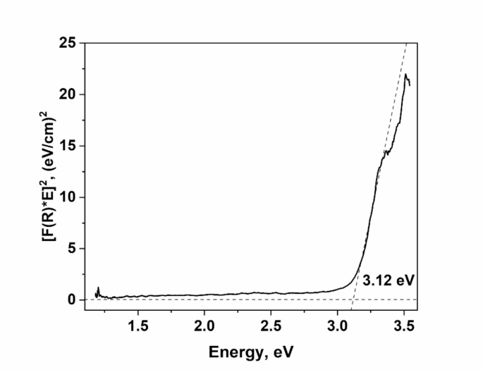
Optical bandgap determined from UV–visible diffuse reflectance spectrum.
According to the modified Kubelka–Munk function, the UV–visible diffuse reflectance spectra show that TiO2 hollow microtetrapods have the bandgap of 3.12 eV. For the purpose of comparison one can note that anatase and rutile phases of TiO2 have a bandgap of around 3.0 eV and 3.2 eV, respectively23, while Zn2Ti3O8 has a calculated bandgap of 3.55 eV 24.
Photoluminescence (PL) measurements were performed to evaluate the presence of defects in the synthesized samples. The PL spectra from Fig. 4a span a broad energy range from 2.0 to 3.5 eV. Notably, as the temperature rises from 10 K to room temperature, the intensity of the high-energy band decreases more significantly than that of the low-energy band.

PL spectra of aero-TiO2 at 10 K and 300 K (a) and the deconvoluted spectrum of PL recorded at 10 K (b).
The deconvolution of the PL spectrum presented in Fig. 4b reveals that it comprises three distinct energy bands: one green band and two violet bands. The green band is centered at 2.5–2.6 eV, while the first and second violet bands are located respectively at 2.9–3.0 eV and 3.15 eV.
One can assign the high-energy emission band at 3.15 eV to near-bandgap transitions, while the violet PL band at 2.9 eV may be associated with an unidentified defect. The green band has previously been linked to the recombination of self-trapped excitons formed from carrier polarons25. It is suggested that oxygen vacancies facilitate effective trapping of carriers or polarons and, simultaneously, the efficient charge separation allows for electron and hole accumulation or trapping at distinct sites, potentially on the surface. Given the extensive surface areas of aeromaterials, one may expect surfaces to play a major role in their photoluminescence behavior.
The kinetics of the tetracycline photodegradation was fitted according to the pseudo-first-order model (Eq. 4):
$$\mathitln\left(\fracC_0C_t\right)=kt,$$
(4)
where, C0 and Ct represent the concentrations of tetracycline in solutions at irradiation time t = 0 min and t, respectively, and k represents the degradation rate (min-1).
Without the photocatalyst, the tetracycline concentration in the solution is not significantly influenced by irradiation with visible or UV light. When aero-TiO2 is added to the solution (Fig. 5e), the concentration of tetracycline decreases by about 75% when irradiated with visible light for 180 min. Upon irradiation with UV light, the photocatalysis process is faster, and the tetracycline concentration decreases by about 90% during 150 min (Fig. 5a). The degradation rates of tetracycline were estimated to be about 0.0064 min-1 and 0.0120 min-1 upon irradiation with visible and UV light, respectively, as shown in Figs. 5b.

Photocatalysis performance of aero-TiO2 for the degradation of tetracycline under visible or UV (a) and under UV light irradiation in the continuous solution flow conditions (c) and their degradation rates (b, d); the schematics of the experimental setup for the photocatalysis tests (e, f).
Previously, Wu et al. have demonstrated that TiO2 P25 nanoparticles with the surface area of about 55 m2/g are able to degrade tetracycline with a ratio of about 0.038 min-1 under 350 nm irradiation, and the photocatalytic efficiency decreases with the increase of the wavelength irradiation source, down to 0.00055 min-1 at 850 nm 26. Despite the differences in testing conditions, these results still can be roughly compared with those from our work. The observed enhanced photocatalytic performance of aero-TiO2 can be attributed to the presence in the nanocomposite structure of Zn2Ti3O8 inclusions decreasing the rate of recombination of photogenerated electron–hole pairs, thus allowing them to reach the photocatalyst surface27,28,29.
Table 1 provides a summary of the existing knowledge on tetracycline photodegradation using various structures of TiO2 photocatalysts, emphasizing the performance of these materials and reaction parameters.
There are three main types of active species which mainly contribute to the photocatalytic degradation of tetracycline, namely ·O2−, ·OH and h+ as was previously observed by other authors37. ·O2− and h+ species have a major role in the photocatalysis under UV and only ·O2− becomes important under visible light irradiation38. The electrons from the valence band are excited to the conduction band, reacting with O2, leading to the formation of ·O2− species, which further react with the adsorbed tetracycline molecules at the material surface, while h+ species directly contribute to the oxidation of tetracycline39.
Previous studies have demonstrated that during photocatalytic oxidation reactions, oxygen vacancy defects in ZnO-TiO2 nanocomposite materials serve as active sites for capturing photoinduced electrons, significantly enhancing photocatalytic efficiency. Additionally, oxygen vacancies facilitate the adsorption of environmental oxygen onto the sample, leading to strong interactions between the photoexcited electrons captured by these vacancies and the adsorbed oxygen molecules40.
The ability to fabricate nanocomposites with precise control opens new pathways for bandgap engineering, enabling alignment of the conduction and valence bands of composite materials with the HOMO and LUMO molecular orbitals of organic compounds targeted for photocatalytic degradation. Under these conditions, photogenerated electrons transfer from the conduction band of one component to that of another, while photogenerated holes similarly move between valence bands. Additionally, transition of the excited electrons from organic molecules to the conduction bands of the nanocomposite components generate reactive species that drive chemical reactions41. Given the substantial specific surface area of the synthesized aeromaterials, it is also likely that surface states play a critical role in modulating the valence band edge, thereby enhancing photocatalytic properties under visible-light irradiation, including those relevant to water splitting.
In the experiment performed under continuous liquid flow conditions using UV light with a density of 3.2 mW/cm2 (see Fig. 5f), the concentration of tetracycline decreases by about 65% during seven hours of irradiation with a degradation rate of 0.0022 min-1 (Fig. 5c and d). After the full degradation of tetracycline, the material was repeatedly used in three more consecutive tests. It was observed that the degradation performance was not influenced, thus demonstrating the reusability of the material. Hence, the 3D shape of our material with 2D features, such as the wall thickness of about 50 nm, makes it suitable for incorporation in active filters for water treatment without the risk of water contamination with the active material.
link


.jpg "IFW Dresden selects Agnitron Agilis 100 MOCVD platform for precursor chemistry and ultra-wide-bandgap materials development")