
Study on the toxic effect of seawater-aged microplastics on Philippine curtain clams

Particle size characterization
A detailed analysis was conducted on the particle size distribution of five types of microplastics (PS, PP, PMMA, PE, and PVC), and the impact of aging on their particle size characteristics was investigated (Table 1). The results indicate that aging exerts varying degrees of influence on the particle size distribution of microplastics. After aging, the particle sizes of PS, PP, and PMMA significantly decreased (P < 0.05), with PS’s DX(90) reducing from 1.189 μm to 1.109 μm, PP’s DX(90) from 0.948 μm to 0.917 μm, and PMMA’s DX(90) slightly from 1.191 μm to 1.177 μm. In contrast, the particle size distribution of PE and PVC expanded after aging, particularly notable in PE, where the DX(90) significantly increased from 0.984 μm to 1.116 μm (P < 0.05). These findings suggest that the effect of aging on particle size distribution are material-specific and vary across microplastic types.
Specific surface area analysis
Based on the data provided in Table 2, we can observe the changes in the specific surface area (m2/g) of different microplastics before and after aging treatment. The aging process generally significantly enhanced the specific surface area of all microplastics (P < 0.001). Specifically, the specific surface area of PS increased by 26.0%, PP by 22.0%, PMMA by 23.9%, PE by 26.0%, and PVC by 24.9%. These changes suggest that the aging process may have led to alterations in the surface morphology of the microplastics, such as an increase in surface roughness or a reorganization of the microstructure, thereby increasing their specific surface area. The statistical significance of particle size characterization and specific surface area analysis indicates that the particle surfaces have been eroded or the particles have undergone fragmentation, resulting in a decrease in particle size and an increase in the exposed surface area. An increase in specific surface area could potentially influence the interactions of microplastics with other substances in the environment, such as enhanced adsorption capacity, which may increase the ecological risks associated with microplastics.
SEM analysis
The SEM analysis of different microplastics samples is presented in Fig. 2. PS microplastics are uniform microspheres with a slightly rough surface and shallow cracks and dents before aging (Fig. 2(a)). The molecular structure of PS contains a benzene ring, which provides stability and results in relatively good aging resistance for PS. After aging, the surface of PS microplastics showed cracks and dents compared to those without aging, and some PS microplastics were cracked and deformed into irregularly shaped fragments (Fig. 2(b)). No significant changes in PP microplastics after aging were observed (Fig. 2(c-d)). PP microplastics are formed through the polymerization and connection of numerous PP fragments. The surface is uneven and irregularly shaped, and due to the molecular structure of PP containing stable carbon-carbon single bonds and carbon-hydrogen bonds, these bonds are not easily broken under conventional conditions. The methyl side group (-CH3) of PP is non-polar, which makes PP highly tolerant to many chemicals and less susceptible to chemical reactions.
PMMA microplastics and PMMA fragments bond and polymerize with each other. Although the arrangement of non-aged PMMA microplastics is disorderly, it is evident that the surface is smooth, with fewer cracks, and the microplastics are relatively full and round(Fig. 2(e)). The molecular structure of PMMA includes stable carbon-carbon single bonds and carbon-hydrogen bonds, which are not easily broken under conventional conditions. Additionally, although the carbonyl group (C = O) of PMMA has a certain polarity, it does not readily undergo chemical reactions under most environmental conditions. However, after continuous exposure to seawater, some PMMA microplastics displayed some cracks (Fig. 2(f)).
The surface of PE microplastics before aging is smooth and uniform, with fewer cracks and a more full appearance(Fig. 2(g)). After aging, the surface of PE microplastics exhibited obvious cracks and depressions (Fig. 2(h)). This may be due to PE being a non-polar hydrocarbon polymer, where oxidation reactions occur, leading to the formation of hydroxyl groups on the molecular chain, reducing the toughness of PE and causing surface cracking.
Unaged PVC microplastics are very round and smooth, with almost no cracks on the surface, appearing very full, and closely resembling spherical shapes(Fig. 2(i)). In contrast to non-aged PVC microplastics, those that have undergone aging show cracks on the surface, and some exhibit significant depressions (Fig. 2(j)). Under the influence of ultraviolet light and high temperatures, PVC may undergo dechlorination reactions, resulting in a decline in material properties, and the appearance of slight cracks and surface depressions.
In summary, PE and PVC samples showed obvious cracks and depressions after aging. PS and PMMA samples exhibited slight cracks and depressions post-aging. No significant aging marks were observed on PP samples, which may be related to the hydrophobicity and insufficient aging time of PP powder. These findings are consistent with those of previous studies50,51,52,53,54.
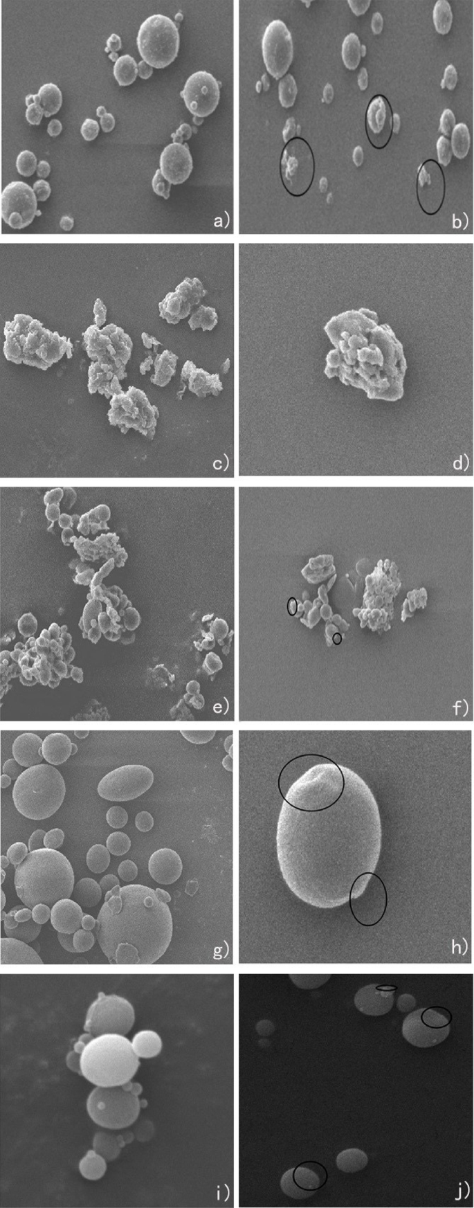
SEM images of microplastics (A: before aging; B: after aging): (a) PS-A; (b) PS-B; (c) PP-A; (d) PP-B; (e) PMMA-A; (f) PMMA-B; (g) PE-A; (h) PE-B; (i) PVC-A; (j) PVC-B.
FTIR analysis before and after aging
Ultraviolet radiation and oxygen exposure are the two main factors that induce the aging process of MPs in aquatic environment. In the FTIR spectra of PS (Figure. 3 (a-b)), the O-H contraction vibration peak appeared in the 3500 cm−155. The absorption peak nearly at 2400 cm−1 is usually associated with the stretching vibration of the carbon-hydrogen (C-H) bond on the benzene ring in PS56.The adsorption band nearly in the 1700 cm ̶1 was attributed to the C = C stretching vibration in the aromatic structure in PS57.
After the aging of PP samples (Figure. 3 (c-d)), superficial signals were observed at 1650–1850 cm−1 and 3250–3600 cm−1, which were determined to be the expansion vibrations of carbonyl groups (i.e., ketones, esters and lactones) and hydroxyl groups (i.e., alcohols and hydroperoxides), respectively58,59. These infrared spectral data showed that the oxidation, hydrolysis, and cracking of PP samples occurred during the aging process, which led to the formation of some new functional groups. Compared with of PMMA samples (Figure. 3 (e)), hydrolytic reaction or oxidation reaction may have led to the formation of a hydroxyl group during the aging process (the characteristic peak at nearly band 3500 cm ̶1 (Figure. 3 (f))60.
After the PE sample aging (Figure. 3(g-h)), a new O-H stretching vibration peak appeared near the band 3500–4000 cm ̶1, indicating that chemical reactions such as oxidation or hydrolysis occurred during the aging process of the PE sample. The formation of new functional groups on the MPs surface, such as vinyl, carbonyl, and hydroxyl/hydroxyl peroxides, resulted in a significant increase in the carbonyl index of MPs, which is consistent with previous studies61.
In the FTIR spectra of PVC (Figure. 3 (i-j)), similar to the FTIR spectra of PS, the O-H contraction vibration peak appeared in the 3500 cm−155. The absorption peak nearly at 2400 cm−1 might exist C = O absorption band in PVC62. The adsorption band nearly in the 1700 cm ̶1 was attributed to the C = O stretching vibration peak appeared in PVC51, and the CH2 deformation appeared in the 1400 cm ̶1 band in PVC63. After aging, the characteristic peaks of the samples did not change, and the shape of the characteristic peaks was basically the same. This indicated that no new functional groups appeared in the aging process of these microplastics, which may have been caused by the short duration and low degradation degree of the aging process of microplastics.

FTIR plots of microplastics (A: before aging, blue line; B: after aging, red line): (a) PS-A; (b) PS-B; (c) PP-A; (d) PP-B; (e) PMMA-A; (f) PMMA-B; (g) PE-A; (h) PE-B; (i) PVC-A; (j) PVC-B.
Exposure experiment results and analysis
Analysis of microplastic lethality
In the 100 mg/L microplastic exposure experiment depicted in Fig. 4, significant disparities in mortality rates over time were observed between unaged and aged microplastics, as well as among different types of microplastics (PS, PMMA, PVC, PE and PP). Adult Philippine clams exposed to non-aged PS, PMMA, and PVC began to show individual deaths after continuous feeding for 72 to 86 h, while those exposed to PE and PP began to die after 100 to 132 h. In the PS, PMMA, and PVC exposure groups, the mortality rate of clams reached 50% between 110 to 120 h, while in the PE group, it was observed at 158 h, and in the PP group, the clam mortality rate remained at 20% from 145 h onwards. Adult Philippine clams exposed to aged PS, PMMA, and PVC began to show individual deaths after continuous feeding for 72 to 86 h, while those exposed to PE and PP began to die after 96 to 120 h. In the PS, PMMA, and PVC exposure groups, the mortality rate of clams reached 50% at 115 to 122 h, while in the PE group, it was observed at 145 h, and in the PP group, the clam mortality rate was 40% at 160 h. In the adult clam group, both non-aged and aged PS, PMMA, and PVC exhibited strong toxicity. Aged PE and PP exhibited stronger toxicity compared to the non-aged group, while aged PS, PMMA, and PVC showed a certain degree of toxicity reduction compared to the non-aged group.
Juvenile Philippine clams exposed to non-aged PS, PMMA, and PVC began to show individual deaths after continuous feeding for 108 to 120 h, while those exposed to PE and PP began to die after 132 to 146 h. In the PS, PMMA, and PVC exposure groups, the mortality rate of clams reached 50% at 145 to 150 h, while at 160 h, the clam mortality rate was 40% in the PE group and 20% in the PP group. Juvenile Philippine clams exposed to aged PS, PMMA, and PVC began to show individual deaths after continuous feeding for 132 to 146 h, while those exposed to PE and PP began to die after 132 h. From the onset of individual deaths until 160 h, in the PS, PMMA, and PVC exposure groups, the mortality rate of clams reached 50% at 155 to 160 h, while in the PE and PP groups, the clam mortality rate was 40% at 160 h. In the juvenile clam group, both non-aged and aged PS, PMMA, and PVC exhibited strong toxicity. Aged PE and PP exhibited stronger toxicity compared to the non-aged group, while aged PS, PMMA, and PVC showed a certain degree of toxicity reduction compared to the non-aged group.
From the above analysis, it is evident that PS, PMMA, and PVC have a stronger toxic effect on both adult and juvenile Philippine clams compared to PE and PP. This might be due to the significantly higher specific surface area of PE and PP before and after aging (Table 1), which allowed them to more effectively adsorb inorganic or organic contaminants in seawater64,65, providing a certain degree of purification to the seawater. Compared to the original microplastics, the toxic effect of aged PS, PMMA, and PVC on both adult and juvenile clams showed a slight reduction, but aged PE and PP exhibited significantly stronger toxicity. This might be due to the formation of new groups in aged PE and PP, such as highly hydrophilic -OH groups, which can induce lipid peroxidation reactions, protein and DNA denaturation in clams66, leading to cellular damage and functional impairment. Although PMMA also formed highly hydrophilic groups during aging, it did not show significant toxicity enhancement, possibly because most PMMA remained hydrophobic after aging67. The mortality rate of juvenile Philippine clams was lower than that of adults under the same conditions, possibly because the daily food intake of juvenile clams was much lower than that of adults, making it more difficult for microplastics to accumulate in their bodies. Additionally, due to their smaller size, juvenile clams might have more difficulty ingesting the gradually floating plastic samples compared to adults. And their overall lower metabolic rate may have influenced how rapidly they accumulated and reacted to toxins.

Mortality of Philippine Curtain Clam in exposure experiments with 100 mg/L concentration of microplastic suspensions: (a) Adult Philippine curtain clams exposed to unaged microplastic; (b) Adult Philippine curtain clams exposed to aged microplastic; (c) Juvenile Philippine curtain clams exposed to unaged microplastic; (d) Juvenile Philippine curtain clams exposed to aged microplastic.
Figure 5 illustrates that in the exposure experiment with a microplastic sample concentration of 500 mg/L, the initiation of mortality in adult Philippine clams exposed to unaged PS, PMMA, and PVC commenced after 60 h of continuous feeding. Conversely, in the exposure experiments involving PE and PP, mortality onset occurred between 84 and 108 h. Within the PS, PMMA, and PVC exposure groups, 50% clam mortality was noted between 110 h and 120 h, whereas in the PE group, it transpired at 142 h, and in the PP group, a 35% mortality rate was observed after 160 h of culture. In adult Philippine clams subjected to aged PS, PMMA, and PVC, mortality was detected after 60 h of continuous feeding, and between 84 and 108 h in the PE and PP exposure experiments. The 50% mortality point for clams in the PS and PMMA exposure groups was reached between 110 h and 118 h, in the PVC group at 132 h, in the PE group at 140 h, and in the PP group, a 35% mortality rate was recorded after 160 h of culture.
Juvenile Philippine clams exposed to unaged PS, PMMA, and PVC exhibited mortality after 84 h of continuous feeding, and between 108 and 120 h in the PE and PP exposure experiments. The 50% mortality thresholds for clams in the PS, PMMA, and PVC exposure groups were achieved at 130 h, 122 h, and 145 h respectively, in the PE group at 150 h, and in the PP group, a 35% mortality rate was documented after 160 h of culture. Similarly, juvenile Philippine clams exposed to aged PS, PMMA, and PVC showed mortality after 84 h of continuous feeding, and between 108 and 120 h in the PE and PP exposure experiments. In the PS, PMMA, and PVC exposure groups, 50% clam mortality was attained at 128 h, 135 h, and 148 h respectively, in the PE group at 150 h, and in the PP group, a 32% mortality rate was recorded after 160 h of culture.
The analysis reveals that, akin to the 100 mg/L exposure group, both juvenile and adult clam populations exposed to unaged and aged PS, PMMA, and PVC exhibited greater toxicity compared to those exposed to PE and PP. Elevating the microplastic sample concentration to 500 mg/L resulted in a marked shift in the mortality timing of Philippine clams, with both the onset of mortality and the time to 50% mortality significantly shortened for both adult and juvenile populations, suggesting a positive correlation between concentration changes and microplastic enrichment in Philippine clams. Nonetheless, there was no discernible difference in mortality rates between the aged and unaged groups across the five materials, possibly due to the high microplastic concentration overshadowing the nuances in plastic types. Future investigations should delve deeper into the specific effects of aging processes on plastic toxicity and assess the contributions of microplastic attributes such as shape, size, and surface roughness to overall toxicity.

Mortality of Philippine Curtain Clam in exposure experiments with 500 mg/L concentration of microplastic suspensions: (a) Adult Philippine curtain clams exposed to unaged microplastic; (b) Adult Philippine curtain clams exposed to aged microplastic; (c) Juvenile Philippine curtain clams exposed to unaged microplastic; (d) Juvenile Philippine curtain clams exposed to aged microplastic.
As depicted in Fig. 6, within the experimental framework involving a 1000 mg/L microplastic suspension, adult Philippine clams subjected to non-aged PS, PMMA, and PVC exhibited initial mortality after sustained ingestion periods ranging from 36 h to 60 h. Conversely, those exposed to PE and PP displayed mortality onset between 72 h and 96 h. Notably, the 50% mortality threshold was reached in the PS, PMMA, and PVC groups at 120 h, 132 h, and 145 h, respectively, with the PE group at 145 h and the PP group at 160 h, registering a mortality rate of 35%. In the context of aged PS, PMMA, and PVC exposure, adult Philippine clams mortality commenced between 48 h and 60 h, with PE and PP exposures initiating mortality between 72 h and 96 h. The 50% mortality mark was achieved in the PS, PMMA, and PVC groups at 138 h, 142 h, and 150 h, respectively, with the PE group at 158 h and the PP group at 160 h, indicating a mortality rate of 40%.
Juvenile Philippine clams exposed to non-aged PS, PMMA, and PVC began to perish after 84 h of continuous feeding, whereas those exposed to PE and PP experienced mortality onset between 108 h and 120 h. The 50% mortality was observed in the PS, PMMA, and PVC groups at 119 h, 132 h, and 144 h, respectively, with the PE group at 144 h and the PP group at 160 h, showing a mortality rate of 35%. In the scenario of aged PS, PMMA, and PVC exposure, juvenile Philippine clams mortality initiation occurred after 84 h of feeding, with PE and PP exposures resulting in mortality onset between 108 h and 120 h. The 50% mortality threshold was met in the PS, PMMA, and PVC groups at 158 h, 140 h, and 148 h, respectively, with the PE group at 150 h and the PP group at 160 h.
The experimental outcomes underscore that non-aged PS, PMMA, and PVC exert a heightened toxicity on both adult and juvenile Philippine clams, manifesting as an earlier mortality onset and a quicker attainment of the 50% mortality threshold. In contrast, PE and PP exhibit a diminished toxicity, with a delayed mortality onset and a protracted timeline for reaching the 50% mortality mark. Aged PS, PMMA, and PVC appear to intensify their toxicity towards adult Philippine clams, evidenced by an accelerated mortality onset and a hastened 50% mortality occurrence. However, the toxicity dynamics of aged PS, PMMA, and PVC towards juveniles are incongruent; certain plastic types (e.g., PMMA) demonstrate a reduction in toxicity, while others (e.g., PS) reveal an escalation in toxicity. This divergence suggests that the aging process may exert varied influences on the toxicity profiles of different plastic types. Under congruent exposure parameters, adult Philippine clams typically exhibit greater susceptibility to plastic toxicity, characterized by an earlier mortality onset and a higher 50% mortality rate. This heightened sensitivity may be attributable to the physiological states, metabolic capacities, and toxic substance tolerances of adults. As the duration of exposure lengthens, the mortality rates of clams across all exposure cohorts escalate, albeit at disparate paces contingent upon the plastic type and aging condition. This variability may be attributable to the bioavailability, bioaccumulation, and specific toxicological mechanisms of plastics on organisms. At a concentration of 1000 mg/L, non-aged PS, PMMA, and PVC demonstrate a more pronounced toxicity towards adult Philippine clams, with an earlier mortality onset and a more rapid attainment of the 50% mortality threshold. This observation implies that the toxicological impacts of plastics may amplify with escalated exposure concentrations.

Mortality of Philippine Curtain Clam in exposure experiments with 1000 mg/L concentration of microplastic suspensions: (a) Adult Philippine curtain clams exposed to unaged microplastic; (b) Adult Philippine curtain clams exposed to aged microplastic; (c) Juvenile Philippine curtain clams exposed to unaged microplastic; (d) Juvenile Philippine curtain clams exposed to aged microplastic.
As shown in Fig. 7, in the exposure experiment with 5000 mg/L microplastic samples, adult Philippine curtain clams exposed to unaged PS, PMMA, and PVC began to die after continuous feeding for 48 h, while individual deaths in the PE and PP exposure experiments started between 72 h and 108 h. The 50% mortality rate in the PS, PMMA, PVC, PE, and PP exposure groups occurred between 142 h and 158 h. Adult Philippine curtain clams exposed to aged PS, PMMA, and PVC began to die after continuous feeding for 36 to 48 h. Individual deaths in the aged PS, PMMA, and PVC exposure groups started between 36 and 48 h, and in the PE and PP exposure experiments, individual deaths occurred between 72 and 108 h. The 50% mortality rate in the PS, PMMA, PVC, PE, and PP exposure groups occurred between 132 h and 155 h.
Juvenile Philippine curtain clams exposed to unaged PS, PMMA, and PVC began to show individual deaths after continuous feeding for 84 h, and in the PE and PP exposure experiments, individual deaths occurred between 108 h and 120 h. Individual deaths were observed at 120 h. The 50% mortality rate in the PS, PMMA, PVC, PE, and PP exposure groups for adult clams occurred between 140 h and 158 h. Philippine curtain clams larvae exposed to aged PS, PMMA, and PVC showed individual deaths after continuous feeding for 84 h, and in the PE and PP exposure experiments, individual deaths occurred between 108 h and 120 h. The mortality rate of the clams increased stepwise from the onset of individual deaths to 160 h. In the PE and PP exposure experiments, the mortality rate of the clams began to rise from 108 to 120 h. The 50% mortality rate in the PS, PMMA, PVC, PE, and PP exposure groups for juvenile clams occurred between 140 h and 158 h.
At a concentration of 5000 mg/L, the onset of mortality in Philippine curtain clams did not advance as it did in other concentration exposure groups, which may suggest that the enrichment rate of microplastics in Philippine curtain clams approached its limit when the concentration was 1000 mg/L. The similar sensitivity of adult and juvenile Philippine curtain clams to microplastics, reaching a 50% mortality rate at similar time points, further indicates that the enrichment rate of microplastics has approached its limit.
The comprehensive analysis indicates that there are differences in the toxicity of various types and conditions (aged versus non-aged) of microplastics to Philippine curtain clams, and factors such as microplastic concentration, clam age, and exposure duration all influence toxicity manifestations.
There were significant differences in the lethality rates of new and aged microplastics at concentrations ranging from 100 to 1000 mg/L (P < 0.05), while there were no significant differences in the values of new and aged microplastics at a concentration of 5000 mg/L. This may be caused by the fact that the excessively high content of microplastics may have masked the original differences between new and aged microplastics. The filtration feeding system and metabolic mechanism of juvenile clams have not been fully developed. At a concentration of 100–500 mg/L, they are more vulnerable to the effects of microplastics. The relatively larger ratio of the surface area to the volume of juveniles makes them have more opportunities to come into contact with microplastics, resulting in significant differences in the mortality rates (P < 0.05). However, at high concentrations of 1000–5000 mg/L, a large amount of microplastics cause serious damage to the physiological functions of both juvenile and adult clams, exceeding the differences in their tolerances, leading to no significant differences in the mortality rates. For the same material at different concentrations, there were certain differences in the toxicity to adult and juvenile clams. Regarding aged microplastics and adult clams, the mortality rate of PP at high concentrations was significantly higher than that at low concentrations. For example, except for the concentration of 5000 mg/L, there were significant differences in the mortality rates among different groups of PE; there was a positive correlation between the concentration of PS and the mortality rate. For juvenile clams, the overall mortality rate caused by aged microplastics was 0%, but there were differences among specific concentrations of some materials.

Mortality of Philippine curtain clams in exposure experiments with 5000 mg/L concentration of microplastic suspensions: (a) Adult Philippine curtain clams exposed to unaged microplastic; (b) Adult Philippine curtain clams exposed to aged microplastic; (c) Juvenile Philippine curtain clams exposed to unaged microplastic; (d) Juvenile Philippine curtain clams exposed to aged microplastic.
The distribution of microplastics within Philippine curtain clams
Upon exposure to stained microplastic suspensions at a concentration of 1000 mg/L for 48 h, the tissue distribution within the Philippine curtain clams was depicted in Figure S4 and S5. In the PMMA exposure group, the soft shell, gills, and digestive glands of the Philippine curtain clams were found to have been stained pink, with the digestive glands exhibiting the deepest coloration, indicating that PMMA had been accumulated predominantly in these tissues, with the highest concentration in the digestive glands (a). The PP exposure group revealed a white soft shell, with minimal stained microplastics having been detected in the gills and digestive glands, suggesting a minor enrichment of PP in these organs (b). Following exposure to PE, the soft shell of the Philippine curtain clams was observed to have taken on a light pink to orange tint, while the gills and digestive glands were notably dark pink, signifying that PE enrichment had been primarily localized to these tissues (c). In the PS exposure group, the soft shell was lightly pink, the gills exhibited a more intense pink, and the digestive glands were comparatively pale, indicating that PS accumulation had occurred across these tissues, with the gills demonstrating the highest concentration (d). The PVC exposure group presented a white soft shell, with the gills and digestive glands having been stained light pink, suggesting a minor presence of PVC in these tissues (e).
Quantitative fluorescence analysis revealed tissue-specific accumulation patterns of microplastics in the Philippine curtain clams, consistent with previous studies11. The distribution characteristics exhibited significant plastic-type dependency and tissue specificity (Figs. 8 and S5). PS showed the highest accumulation in gill tissue (6.25 µg/g, 28.92%), significantly exceeding other tissues, likely due to the affinity of its benzene ring structure for gill mucosal surfaces, while maintaining substantial levels in the digestive gland (5.67 µg/g, 26.24%). PMMA demonstrated predominant enrichment in the digestive gland (6.6 µg/g, 35.00%, P < 0.05 versus other tissues), aligning with its hydrophilic properties that facilitate filter-feeding uptake. PE exhibited a dual accumulation pattern in both gills (4.68 µg/g, 32.03%) and digestive gland (4.39 µg/g, 30.04%), with significant inter-tissue differences (P < 0.05) reflecting its moderate hydrophobicity-mediated trans-tissue transport. PVC showed comparable distribution between gills (3.82 µg/g, 35.03%) and digestive gland (3.93 µg/g, 36.03%, P > 0.05), whereas polypropylene (PP) accumulated predominantly in the digestive gland (2.15 µg/g, 38.91%) and gills (2.03 µg/g, 36.74%), both exhibiting limited tissue penetration due to high hydrophobicity. Notably, distinct tissue distribution patterns emerged among plastic types ( P < 0.05): PMMA and PP showed the highest relative proportions in the digestive gland (35.00% and 38.91%, respectively), while PS and PVC maintained balanced gill-digestive gland distributions, attributable to synergistic effects between physicochemical properties (e.g., hydrophilicity/hydrophobicity, functional groups) and biological uptake pathways (filter-feeding, respiration).
The results demonstrate that microplastic distribution is jointly regulated by physicochemical properties and uptake mechanisms68. PMMA’s hydrophilicity promotes digestive gland enrichment via filter-feeding; PE’s moderate hydrophobicity enables gill capture followed by digestive gland transfer; PS’s benzene ring enhances gill adhesion; while Pp and PVC’s high hydrophobicity restricts tissue permeation, with PVC’s density further reducing bioavailability. Crucially, all tested microplastics were detected in gills, confirming respiration as a major uptake route, while digestive gland dominance underscores filter-feeding’s pivotal role. These findings systematically elucidate microplastic translocation and accumulation mechanisms in aquatic filter-feeders, providing critical scientific basis for ecological risk assessment.

Fluorescence intensity of microplastics distribution in the Philippine curtain clams. The different letters inside the cumulative bars represent the significant differences in the proportion of fluorescent intensities between different organs.The letters following the hyphen (-) represent significant differences in the proportion of fluorescent intensities among different plastics within the tissue.
link


.jpg "IFW Dresden selects Agnitron Agilis 100 MOCVD platform for precursor chemistry and ultra-wide-bandgap materials development")